

3D dental imaging—also known as cone-beam CT (CBCT) scanning—is a revolutionary X-ray technology that creates a three-dimensional view of your teeth, jaws, and surrounding anatomy. Instead of a flat 2D picture, a CBCT scan rotates a cone-shaped X-ray beam around your head and captures hundreds of images. A computer then stitches these “slices” into a detailed 3D model of your mouth. This means we can see the exact shape and depth of structures (like nerve canals or tooth roots) that would be invisible on a standard X-ray. For example, Arlington Dental uses a state-of-the-art Carestream Planmeca CS 9300 system, which provides “crystal-clear” 3D images with much lower radiation exposure than conventional CT scans. By capturing all angles in one quick scan, 3D imaging helps our dentists diagnose issues and plan treatment far more accurately than ever before.
Modern dental imaging devices (like the X-ray unit shown above) have evolved from simple film X-rays to advanced 3D scanners. Today’s CBCT machines allow Arlington-area dentists to capture an on-screen 3D model of your jaws. As the American College of Radiology explains, dental cone-beam CT produces 3D images of teeth, soft tissues, nerve pathways and bone in a single scan, providing much more information than regular bitewing or panoramic X-rays. In practice, this means we can identify small fractures, hidden canals, and early-stage diseases without guesswork.
How 3D Imaging (Cone-Beam CT) Works
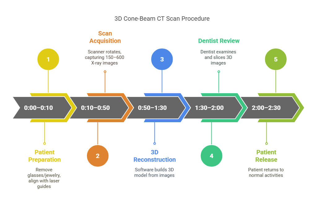
Rather than a quick snapshot, a 3D CBCT scan is more like creating a “flip book” of X-ray slices. The patient sits or stands very still while the cone-beam scanner rotates around the head, capturing up to 150–600 individual X-ray images in under a minute. These images are then reconstructed by software into a manipulable 3D model. In our Arlington office, the process is straightforward and comfortable for patients. You’ll be asked to remove glasses or jewelry, and a staff member will position the open-ring scanner around your head. As shown below, red laser guides help align you properly before the scan. Then the machine quietly spins for only 10–40 seconds while it captures the data.
During a 3D cone-beam scan, a lightweight halo frame and lasers are used to align your head. Once positioned, the scanner rotates around you for just seconds (you may hear a soft whirring). The procedure is completely painless – there are no needles or discomfort – and you simply hold still while the images are taken. Immediately after the scan, powerful software reconstructs the 3D model on-screen. In fact, Arlington Dental notes that dentists can “inspect the images on screen, zooming in on any area and taking virtual ‘slices’ as needed”. This lets us examine your teeth and jaws from every angle without any additional X-rays. The whole visit often takes just a few minutes, and you can return to normal activities right away.
Benefits of 3D Dental Imaging
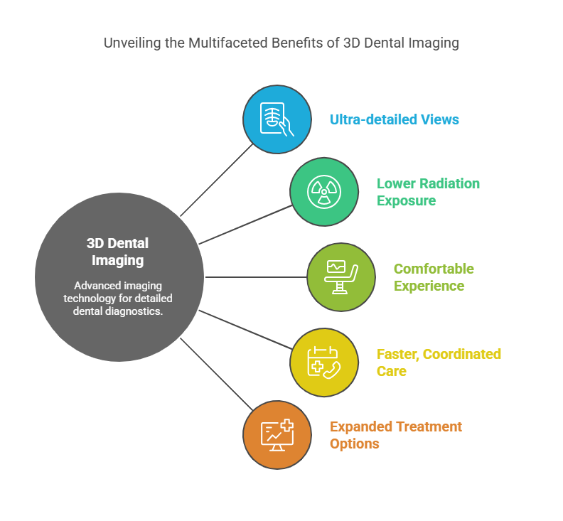
3D imaging offers clear advantages over traditional X-rays. In general, cone-beam CT scans deliver higher diagnostic value with less radiation and greater comfort. For instance, Arlington Dental highlights these benefits:
-
Ultra-detailed views: 3D scans capture teeth and jaw anatomy in “unprecedented detail,” so we see fine structures (tiny root canals, bone defects, nerve paths) much more clearly than on 2D films. This high resolution can uncover problems earlier and guide more precise treatment.
-
Lower radiation exposure: Although a cone-beam scan uses more X-ray dose than a single bitewing, it focuses only on the needed area. Modern CBCT units like the CS 9300 minimize exposure by selecting a small “field of view” to target your area of concern. In practice, a dental CBCT scan usually imparts only a few days’ worth of background radiation – far less than a hospital CT. We follow the ALARA principle (“As Low As Reasonably Achievable”) to keep doses minimal.
-
Comfortable experience: CBCT machines are open design, so you don’t lie in a claustrophobic tunnel. You can sit or stand during the scan. There is no pain or anesthesia needed, unlike some older imaging methods. In fact, most patients find the experience quick and easy.
-
Faster, coordinated care: Because 3D scans capture all angles in one go, you often leave with answers immediately. There’s no need for a separate radiology appointment. Our dentist can review the images with you on the same visit, helping you understand any issues right away. Additionally, if multiple specialists are involved in your care, 3D images can be easily shared with your orthodontist, oral surgeon or lab, improving coordination.
-
Expanded treatment options: Access to 3D imaging allows dentists to diagnose and treat conditions they couldn’t before. We can perform a wider range of in-office procedures (from complex extractions to implant placement) with confidence. In short, 3D scans support more efficient and effective care.
Common Uses of 3D Imaging in Arlington Dentistry
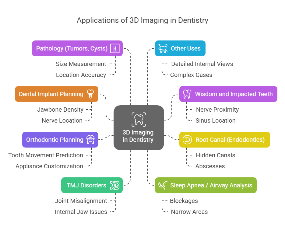
Dentists and specialists use 3D CBCT scans for many advanced procedures. In our Arlington practice, some of the most frequent applications include:
-
Dental implant planning: A CBCT scan shows the exact shape and density of your jawbone, and the precise location of nerves and sinuses. This 3D roadmap lets us place implants with millimeter accuracy. As Arlington Dental notes, detailed CBCT images ensure we “avoid nerves, sinuses and areas of low bone density” and choose the ideal implant position.
-
Wisdom and impacted teeth: Impacted teeth can be close to nerves or roots of adjacent teeth. A 3D scan reveals the exact position of a wisdom tooth or other impacted tooth relative to your nerves and sinus. This helps us plan a safe extraction or exposure with minimal risk.
-
Root canal (endodontics): Complex root canal cases often involve hidden canals or cracks that a normal X-ray might miss. A CBCT scan “reveals hidden canals and abscesses,” improving the success rate of root canals. In endodontics, seeing the tooth’s full 3D anatomy can make the difference in diagnosing a tough problem.
-
Orthodontic planning: Braces and clear aligners work by moving teeth through bone. A 3D scan shows the current tooth and jaw relationship in all dimensions, allowing precise planning of tooth movements. Orthodontists use CBCT to predict how teeth will shift, and to customize appliances for your mouth.
-
TMJ (jaw joint) disorders: If you have jaw pain or clicking, 3D imaging gives detailed coronal, sagittal and axial views of your temporomandibular joints. We can spot joint misalignment, arthritis, or internal jaw issues that wouldn’t appear on a 2D film.
-
Sleep apnea / airway analysis: CBCT can even visualize the airway and sinus passages. For patients with suspected sleep apnea or nasal obstructions, a 3D scan helps identify any blockages or narrow areas in the throat and nose.
-
Pathology (tumors, cysts): Any unusual lesion, cyst or bony abnormality in the jaw can be fully assessed with 3D imaging. It lets us measure the size and location of growths accurately and plan appropriate treatment.
-
Other uses: Virtually any time we need a highly detailed internal view of the teeth, bone or facial anatomy, CBCT is useful. In summary, whenever a flat X-ray isn’t enough, 3D imaging is invaluable.
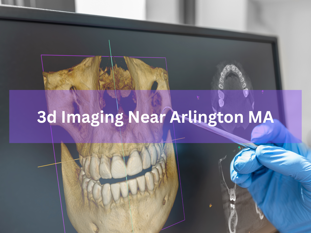
3D imaging can reveal problems that 2D X-rays alone might miss. For example, the detailed scan above (with the blue-gloved hand pointing) shows tooth roots and surrounding bone clearly. Such high-resolution images help detect decay between teeth, root fractures, or even early cysts around an impacted tooth – all before symptoms occur.
3D Imaging at Arlington Dental
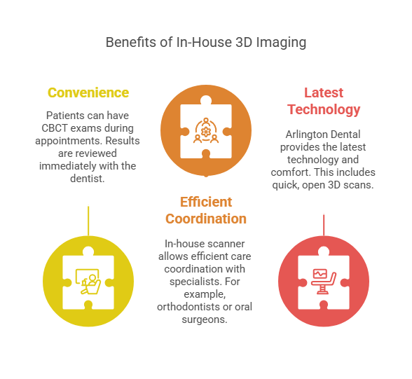
At Arlington Dental’s office in Arlington, MA (43 Broadway), we’ve invested in an in-house 3D imaging system so our patients don’t have to visit an outside imaging center. Our CS 9300 cone-beam CT unit lets us capture scans right here on site. This means you can have the CBCT exam during your appointment, and review the results immediately with our dentist. Having the scanner in-house also allows us to coordinate care efficiently with any specialists you may need (for example, an orthodontist or oral surgeon). We strive to provide every Arlington patient with the latest technology and comfort – which includes quick, open 3D scans delivered directly from our practice.
What to Expect During Your 3D Dental Scan
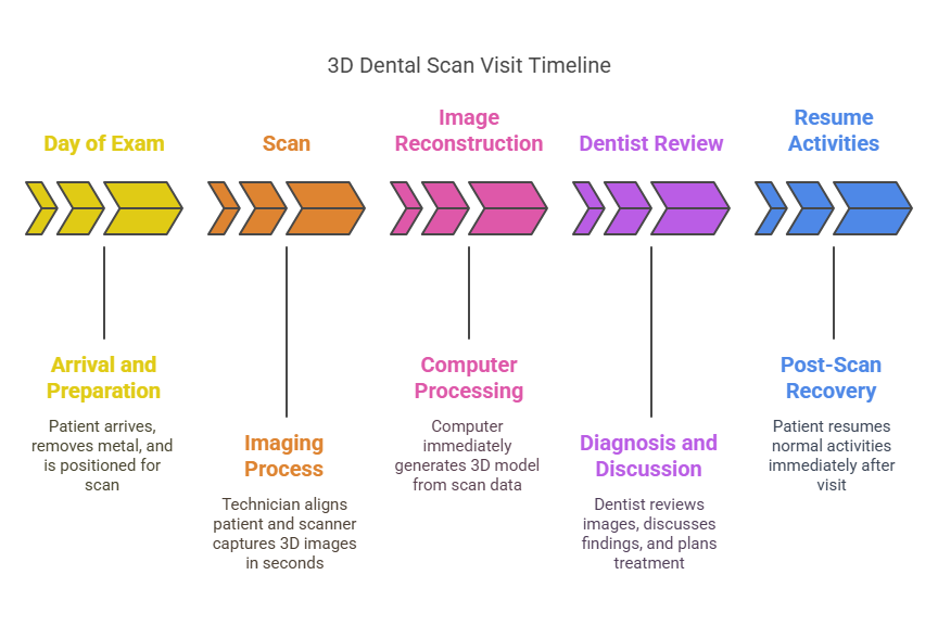
Preparation for a 3D scan is minimal. Wear comfortable clothing and remove any metal accessories (glasses, jewelry) that could block X-rays. On the day of your exam, our technician will seat you or have you stand beside the CBCT machine and position the head alignment frame. You’ll see red laser light lines projected on your face to ensure precise placement (as shown above). Then the scan takes place: you simply hold very still while the unit rotates. The actual scan time is only about 10–20 seconds. You will hear a gentle whirring, but you won’t feel a thing – the process is non-invasive and painless.
Once the scan is complete, the computer immediately reconstructs the 3D model. Within moments, our dentist can view the full volumetric image on-screen, zoom into any area, and even take “virtual slices” through the 3D data. The entire 3D imaging visit (including setup and scan) usually takes only a few minutes longer than a regular X-ray appointment. Afterward, you can eat, drink and resume normal activities right away. Our goal is to make the experience as quick and comfortable as possible. As Arlington Dental notes, this modern process lets you “leave the appointment with a complete treatment plan,” since diagnosis and planning can happen immediately. For those interested in the clinical applications of 3D imaging in orthodontics, particularly in treating complex cases like Class III malocclusions, the American Association of Orthodontists offers an insightful educational resource on 3D Imaging in Class III Orthopedic Treatment.
The graphic above outlines a typical 3D scan visit timeline. On the day of exam you come in (yellow), prepare and get positioned, and the scanner takes the images. The scan (orange) is followed by image reconstruction on the computer (pink). Then the dentist reviews the images and discusses them with you (purple), and finally you go home and resume normal activities (blue). As you can see, the entire sequence from start to finish is brief and efficient.
Safety and Radiation
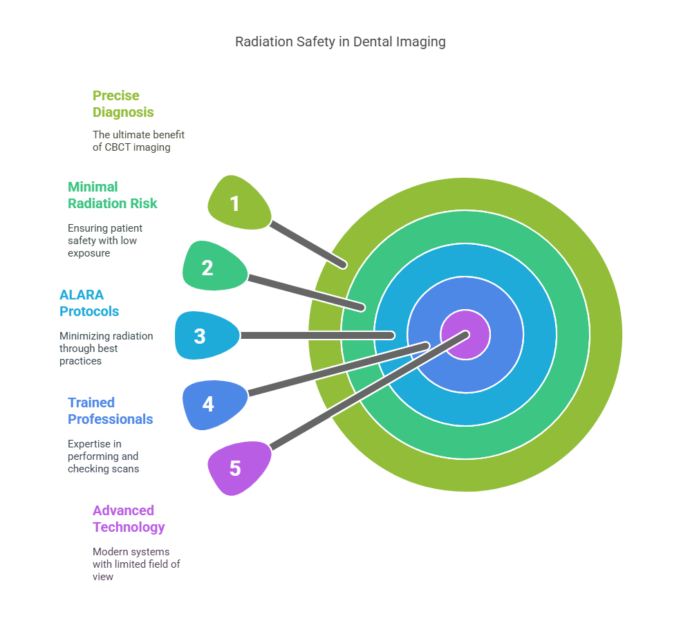
Patient safety is our top priority during any imaging procedure. While cone-beam CT does use X-rays, the radiation dose is very low when used properly. Unlike a full medical CT scan, a dental CBCT focuses on a limited area (just your mouth and jaw). Modern systems like ours allow us to choose a small “field of view” so only the region of interest is exposed. By following ALARA protocols (As Low As Reasonably Achievable), we minimize every scan. For perspective, the radiation from a single dental CBCT is typically equivalent to just a few days of natural background radiation – much less than most people receive on a cross-country airplane flight, and far lower than a hospital-grade head CT. In fact, Boston University has reported that their dental CBCT delivers “substantially less radiation” than a standard medical CT. We also use lead aprons/thyroid collars when indicated and reserve CBCT for cases where the additional detail is needed.
In practice, CBCT’s detailed images allow us to shorten or avoid more invasive procedures, which can be safer overall. As the American Dental Association notes, dental imaging accounts for a very small fraction of total medical radiation exposure. At Arlington Dental, every 3D scan is performed by trained professionals and checked for necessity. Rest assured that when we recommend 3D imaging for your care, the benefit of a precise diagnosis far outweighs the minimal radiation risk.
Cost and Insurance
Many patients wonder about the cost of advanced imaging. Insurance coverage for CBCT scans varies by plan. Generally, dental or medical insurers will cover a 3D scan when it is medically necessary – for example, if it’s needed for dental implant planning, wisdom tooth surgery, or diagnosing a complex pathology. Some plans classify CBCT as an “alternate radiographic” exam. At Arlington Dental, we will help you check your benefits in advance. In many cases, the imaging fee can be included with your treatment (such as bundling the scan with implant placement). Even if your plan doesn’t fully cover it, we offer flexible financing options to make the technology accessible. Ultimately, we believe the precision and improved outcomes gained from 3D imaging are well worth the investment in your dental health.
People Also Ask
Q: What is 3D dental imaging? A: 3D dental imaging, or cone-beam CT, is a specialized X-ray that produces a three-dimensional view of your teeth, jaws and face in a single scan. It captures many X-ray “slices” around the head and combines them into a virtual 3D model. This provides more detailed information than flat 2D X-rays, revealing depth, hidden anatomy, and exact relationships of structures. In short, it lets dentists see things they couldn’t on a regular X-ray.
Q: How long does a 3D dental scan take? A: Very little time. Modern cone-beam CT units are fast. The scanner rotates around your head in just 10–20 seconds and captures all the needed data. Including setup and positioning, the entire visit usually takes only a few extra minutes longer than a routine X-ray appointment.
Q: Is a 3D dental scan painful? A: Not at all. A 3D scan is completely painless and non-invasive. You simply sit or stand still while the machine moves around you. There are no needles, no physical discomfort, and no injections. You will hear the equipment move (a quiet whirring), but otherwise feel nothing.
Q: How much radiation does a 3D dental X-ray use? A: Cone-beam CT uses more radiation than a single bitewing X-ray but significantly less than a medical CT scan. For example, our CS 9300 unit delivers crystal-clear 3D images while minimizing exposure to radiation. In real terms, a full 3D dental scan usually exposes you to about the same amount of radiation you’d get from a couple days of natural background exposure. This dose is far lower than a hospital CT scan of the head, and modern protocols ensure it stays as low as possible.
Q: Why do dentists use 3D imaging? A: Because it shows hidden details that 2D X-rays can’t. For instance, 3D scans can pinpoint the exact location of nerves, measure jawbone density for implants, or spot issues in the sinus and airway. This leads to safer, more effective treatment plans. In short, 3D imaging increases diagnostic confidence and helps avoid surprises during procedures.
Q: Where can I get a 3D dental scan in Arlington, MA? A: Our practice at Arlington Dental (43 Broadway, Arlington, MA) has an in-house 3D cone-beam CT scanner. You can get the scan right here in our office during your appointment. Because we have our own CBCT unit, there’s no need to visit an outside facility – it can all be done in one place for your convenience.
Q: How does 3D imaging benefit dental implants? A: Before placing an implant, it’s crucial to know exactly how much bone you have and where vital structures lie. A 3D CBCT scan provides a virtual model of your jawbone in 360 degrees, allowing precise implant planning. The scan shows us the ideal implant size, location and angle, helping avoid nerves or sinuses. This precision minimizes surgical risks (like nerve injury) and often leads to better healing and success on the first try.
Q: Are 3D dental scans covered by insurance? A: Insurance coverage varies. Many dental and medical plans will cover a 3D scan if it’s deemed necessary for a covered treatment – for example, implants, oral surgery or pathology. Sometimes CBCT is billed as an “alternate radiographic” exam. At Arlington Dental, we submit the claim for you and can discuss your benefits. Even if coverage is limited, we offer payment options. Our team is happy to help you understand the costs based on your specific insurance.
Q: Can children have 3D dental imaging? A: Yes, if it’s needed for their care. Because children’s developing tissues are more sensitive, we only use CBCT for pediatric patients when it’s truly warranted (for example, complex orthodontic cases or surgical planning). Otherwise, routine dental X-rays (with very low dose) are preferred for kids. In any case, our facility follows strict safety protocols and would only recommend a scan for a child if the benefit of having the detailed 3D information clearly outweighs any risk.
Q: How is cone-beam CT different from a regular (medical) CT? A: Cone-beam CT is specially designed for dental use. It uses a cone-shaped X-ray beam and captures a full 3D jaw image in one quick rotation. In contrast, a medical CT makes many parallel slices and usually scans the entire head or body. The cone-beam scan produces very similar quality images for teeth and jaws but with a much smaller machine, lower radiation dose and lower cost. In short, CBCT gives dentists CT-like detail in an affordable, office-friendly format.
Frequently Asked Questions (FAQ)
Is 3D dental imaging better than ordinary X-rays? Yes. Unlike flat X-rays, 3D imaging provides depth and can reveal hidden anatomy. It often detects issues (like fractures or additional canals) that a 2D image might miss.
How should I prepare for a 3D scan? Preparation is easy: wear normal clothes and remove glasses, earrings, or metal jewelry. Apart from that, no special prep (like fasting) is needed. You may be asked to wear a lead apron for safety, just as with any X-ray.
Will I need a referral to get a CBCT scan? At our office, no referral is needed. Because we have the equipment in-house, our dentist can order the scan directly during your visit if indicated.
Can pregnant patients have 3D dental imaging? Generally, we avoid any X-rays during pregnancy unless absolutely necessary. If a scan is urgent, extra precautions (like shielding) are used, but often we postpone elective imaging until after pregnancy.
Does sedation or numbing help with the scan? No sedation or numbing is required for a CBCT scan. It is non-invasive and takes only a few seconds. You just need to stay still; the machine does all the work.
What does a 3D scan show that an X-ray doesn’t? A 3D scan shows the exact 3D shape of teeth, bone, nerves and sinuses. For example, it can reveal the thickness of bone, hidden cysts, or the true path of a root canal – details that overlap in an X-ray image.
How much does a 3D dental scan cost? Costs vary by case and insurance. Many times the scan fee is included with a treatment (like implants). If needed, our office can provide estimates or finance plans. We can also tell you what portion, if any, your insurance is likely to cover.
Is cone-beam CT painful or uncomfortable? Not at all. It’s usually more comfortable than sitting for a long exam or having a tooth extracted. You simply remain still while the machine rotates; there is no pain involved.
Who benefits most from 3D dental imaging? Patients undergoing complex procedures (implants, root canals, extractions) or those with unexplained dental issues benefit greatly. Also anyone needing a second opinion on a difficult case. Essentially, anyone who wants the most thorough diagnostics available can consider it.
How often should I get 3D dental scans? Only when recommended by your dentist. Unlike routine bitewings or panoramics, 3D scans are used selectively for specific diagnostic needs. We use them only when the added information will significantly impact your care.
Each of the answers above provides quick, useful information. If you have more questions about 3D dental imaging in Arlington, MA, our friendly team at Arlington Dental is happy to explain further and guide you to the best options for your care.
Conclusion: The Future of Precision Dentistry
3D dental imaging is no longer a luxury—it’s becoming the new standard in high-quality dental care. At Arlington Dental, we are proud to offer this advanced technology right here in our office, allowing us to diagnose, plan, and treat with unmatched accuracy and efficiency. Whether you need a dental implant, evaluation for jaw pain, or a detailed orthodontic workup, cone-beam CT gives us the insight to provide safer and more predictable outcomes. Our commitment to innovation means we can deliver modern care with minimal discomfort and maximum precision—so you leave each visit confident in your treatment plan. If you’re in Arlington, MA and want to experience the benefits of 3D dental imaging for yourself, schedule an appointment today and see how this breakthrough technology is transforming smiles. Top Arlington MA dentist available at Arlington Dental, 43 Broadway, Arlington, MA 02474. (781) 641-0500.